Blumenthal EZ, Horani A, Sasikumar R, Garudadri C, Udaykumar A, Thomas R.
Ophthalmic Surg Lasers Imaging. 2006 May-Jun;37(3):218-23.
Department of Ophthalmology, Hebrew University-Hadassah Medical Center, Jerusalem, Israel.
ABSTRACT:
BACKGROUND AND OBJECTIVE: To correlate structure and function in eyes with end-stage glaucoma.
PATIENTS AND METHODS: Fifty-six eyes of 48 patients with glaucoma presenting with end-stage glaucoma underwent scanning laser polarimetry (SLP) imaging using a commercially available GDx-variable corneal compensator unit (GDx-VCC; Laser Diagnostics Technologies, Inc., San Diego, CA). End-stage glaucoma was defined by both disc appearance and standard automated perimetry visual field criteria. Standard automated perimetry parameters included: mean deviation, pattern standard deviation, and total deviation plot. GDx parameters included: TSNIT average, superior average, inferior average, TSNIT standard deviation, and nerve fiber indicator.
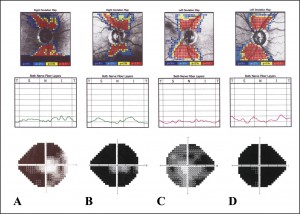
RESULTS: The visual field mean deviation was -26.75 +/- 3.50 dB. The remaining retinal nerve fiber layer measured in this group of eyes was: TSNIT average, 29.76 +/- 5.81 microm; superior average, 30.76 +/- 6.25 microm; and inferior average, 31.14 +/- 7.20 microm. A low structure-function correlation was found when analyzing separately the superior and inferior hemifields (R2 = 0.00001, R2 = 0.0016, respectively).
CONCLUSIONS: In eyes with end-stage glaucoma, very thin but existing retinal nerve fiber layer is found on SLP. Such values rarely dropped below 10 to 20 microm. A flattening of the GDx TSNIT pattern was seen, and the correlation between structure and function was not evident.
Figure from this article:

